

OEM Transcatheter Aortic Valve: Revolutionizing Minimally Invasive Heart Care
2026-11-16
Heart valve replacement has long meant cracking open the chest. But a quiet revolution is underway in interventional cardiology, and it fits through an artery. OEM transcatheter aortic valves are reshaping what’s possible, turning high-risk open-heart procedures into controlled, catheter-based moments of precision. Behind some of the boldest advances is INT Medical, a partner that doesn’t just manufacture—it co-engineers devices trusted when every heartbeat counts. Let’s step inside the technology, the partnerships, and the unspoken question this innovation raises: How much more can we fix without ever opening a chest?
How a Foldable Valve Reaches the Heart Without Open Surgery
A foldable valve is carried through the body’s vascular highways using a slim delivery catheter. The journey usually begins at a small incision in the groin or chest, where the catheter enters a major artery or vein. Compressed to just a few millimeters, the valve slips through these pathways until it reaches the heart chambers, guided by real-time imaging that lets the team watch every twist and turn.
Once the new valve sits inside the failing native valve, the real work starts. A balloon inside the catheter inflates—or sometimes the valve’s own frame springs outward—expanding the device until it pushes the old, calcified leaflets aside. The foldable structure locks into place and immediately takes over the job of directing blood flow. Patients are usually awake, the heart never stopped, and the chest never cracked open.
The beauty of this approach isn’t just in the smaller scar. Because the body isn’t subjected to the trauma of a major incision, recovery can shrink from months to days. Many people go home within 48 hours, breathing easier and regaining energy that had been stolen by a worn-out valve. It’s a design that turns a complex cardiac repair into a procedure that feels almost like a long diagnostic session.
The Quiet Engineering Feat Inside a Collapsed Valve
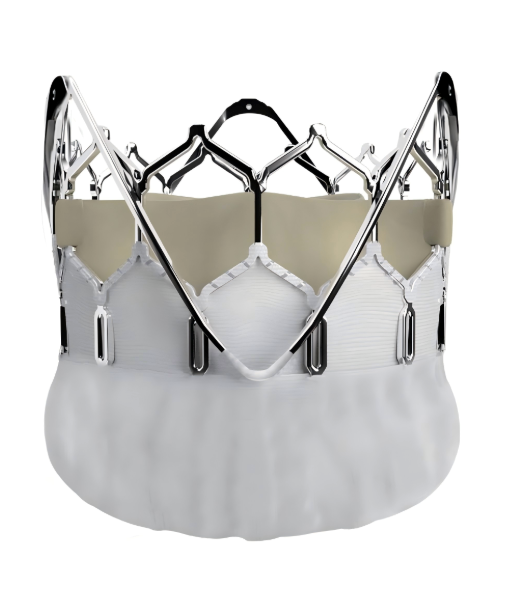
Most people never think about a valve until it fails, and when it does, the failure is rarely quiet. But inside a collapsed valve lies a hidden lesson in material memory and pressure balance. The seemingly inert metal components have been quietly flexing for years, enduring micro-deformations that accumulate like fatigue in a long-distance runner. When the valve finally gives way, it is not a sudden betrayal but the inevitable resolution of countless silent negotiations between stress and structure.
What makes this collapse a quiet engineering feat is not the failure itself but the story told by the fracture surfaces. Under an electron microscope, the twisted grain structures reveal the valve’s entire operational history—every thermal cycle, every pressure spike, every vibration damped by its own resilience. Engineers often see such valves as passive components, but in reality, each one holds a dynamic record of the system it once served. The collapse becomes a final, eloquent statement about the limits of predictability in closed systems.
This hidden language of collapse reshapes how we approach design margins. Rather than simply strengthening parts, the insight from a collapsed valve points toward more adaptive materials that can signal their degradation before failure. It is a quiet reminder that the most meaningful engineering breakthroughs sometimes emerge not from flawless performance but from the quiet, forensic reading of what went wrong.
From Femoral Artery to Aortic Annulus: Mapping the Delivery Route
Navigating from the femoral artery up to the aortic annulus is a journey that demands more than just anatomical knowledge—it requires a real-time appreciation of the twists, turns, and subtle variances that make each patient's vascular tree unique. Think of this route not as a rigid blueprint but as a living map, where the iliac bifurcation, the arch's curvature, and the calcified segments act as landmarks that can either guide the delivery system smoothly or challenge its passage. Clinicians often rely on a mental overlay of preoperative imaging and intraoperative tactile feedback, constantly adjusting their approach as the catheter tip encounters resistance or confirms a familiar bend.
The mapping process itself is iterative. Starting from the common femoral entry, the wire and catheter traverse the external iliac, common iliac, and then navigate the aortic bifurcation—a critical point where tortuosity or plaque can redirect the path unpredictably. Once in the abdominal aorta, the journey upward passes by the renal and visceral branches, which must be respected to avoid inadvertent cannulation. The aortic arch presents perhaps the most complex segment: the curve toward the annulus often requires a supportive guidewire shape, and the final descent toward the aortic valve calls for precise control to prevent leaflet damage or annular disruption. Each section of the route holds its own set of potential hang-ups, and seasoned operators learn to read the wire’s behavior as a silent narrative of the vasculature ahead.
Beyond mere navigation, this route serves as the conduit for delivering therapeutic devices—be it a transcatheter heart valve, a balloon-expandable stent, or a closure device. Understanding the delivery route means respecting the cumulative impact of friction, vessel angulation, and diameter changes on the delivery system’s performance. A smooth journey from the femoral artery to the annulus relies on the operator's ability to predict how the anatomy will guide—or obstruct—the hardware. Ultimately, mapping this path isn’t just about reaching the destination; it’s about doing so in a way that preserves vessel integrity and sets up the precise, stable positioning needed for a successful intervention.
Why Tissue Durability Matters More Than Frame Design
Most buyers instinctively judge a sofa by its silhouette or how solid the frame feels when they nudge it with a knee. That’s a distraction. A sturdy frame is meaningless if the fabric sitting on top of it starts pilling, fading, or thinning out after a year of real life. Tissue—meaning the textile that actually meets your skin—takes every ounce of daily abuse: sunlight, pet claws, skin oils, spilled coffee. It’s the barrier that preserves the piece’s dignity long after the novelty of its shape wears off.
Look at it this way: no one ever gets rid of a couch because the internal joinery suddenly failed while they were watching television. They retire it when the upholstery turns shabby. A well-chosen, high-rub-count fabric can make even a mediocre frame last a decade longer in a home simply because people don’t feel the need to replace it. Durability is emotional, not just physical. When the textile holds up, the whole sofa stays loved.
Frame design is about the first impression; tissue durability is about the relationship. You can have elegantly tapered legs and a perfectly angled backrest, but once the weave starts to bag or the color washes out unevenly, that initial romance is over. The fabric’s resilience determines whether the sofa remains a comfort zone or becomes an eyesore you’re constantly apologizing for. In the long run, your skin and your daily routines care far more about what’s touching them than about what’s hidden under the foam.
Reading Patient Anatomy Through Pre-Procedural Imaging
Reading Patient Anatomy Through Pre-Procedural ImagingReal-World Rhythm: Recovery Patterns After Valve Deployment
When a new valve settles into place, the body doesn’t simply restart—it learns a different tempo. The first few days are a quiet negotiation between circulation and compensation. Breathing deepens without the old strain. Someone who used to pause midway up a staircase now walks the cardiology ward halls with a steadiness that feels almost foreign. This shift isn’t dramatic; it’s a gradual recalibration, often described as waking up to a rhythm that finally fits.
Then comes the messy middle, where real-world recovery rarely obeys discharge pamphlets. Some patients vault forward in weeks, crediting pre-procedure resolve or just plain luck. Others drift through a stretch of ectopic beats and low-grade uncertainty as the heart’s electrical network adapts to a reshaped chamber. It’s not a straight line—more like a dance where ventricle, valve, and vascular tree learn each other’s moves. Fatigue still visits, but it’s a different tired, one that doesn’t linger as long.
By the third or fourth month, the pattern turns personal. A retired teacher bends to pull weeds without needing a break. A weekend golfer rediscovers his back nine without the familiar mid-round fade. These aren’t metrics you’ll find on an echocardiogram; they’re the quiet signatures of recovery, written in daily routines reclaimed and breaths taken for granted. The valve doesn’t just function—it becomes part of a lived rhythm, steady and unannounced.
FAQ
It refers to a valve manufactured by a specialized partner but branded and distributed by another company under its own label. While the core technology mirrors a standard transcatheter aortic valve—designed for implantation without open-heart surgery—the OEM model allows medical device firms to bring proven designs to market faster, often tailoring the delivery system or leaflet material to specific clinical needs without reinventing the entire device.
It eliminates the need for a sternotomy and cardiopulmonary bypass. Instead, the replacement valve is compressed onto a catheter, threaded through an artery (usually in the leg), and expanded inside the diseased native valve. This dramatically reduces recovery time, lowers the risk of infection, and makes aortic valve replacement possible for elderly or frail patients who were previously deemed inoperable.
Most procedures are performed under conscious sedation rather than general anesthesia. The patient is usually awake but comfortable. Post-procedure, many are sitting up within hours and discharged in two to four days, compared to a week or longer with open surgery. The immediate relief from symptoms like shortness of breath and chest tightness can be quite striking.
Real-time imaging is critical. A multimodality approach—combining fluoroscopy and transesophageal echocardiography—guides every step, from crossing the narrowed aortic valve to precisely positioning and deploying the new valve. Pre-procedural CT scans are also essential for sizing the valve and mapping the access route, helping to avoid complications like paravalvular leakage or coronary obstruction.
Long-term data up to 8–10 years now shows comparable durability for many transcatheter valves, with low rates of structural deterioration. Innovations in leaflet design—whether bovine pericardium treated with advanced anti-calcification methods or novel polymer materials—continue to push projected lifespans further. Regular echocardiographic follow-up is routine to monitor function, just as it would be for a surgical bioprosthesis.
It often leads to more competitive pricing and reliable supply chains. By procuring valves through established OEM partnerships, hospitals can access clinically proven designs without the premium of a big-name brand. This can accelerate adoption in cost-sensitive environments, expand patient access to minimally invasive care, and still maintain rigorous quality and traceability standards.
Conclusion
The OEM transcatheter aortic valve redefines cardiac intervention by merging delicate foldable mechanics with a delivery journey that starts in the groin and ends at the aortic annulus. Compressed within a slender catheter, the valve travels retrograde through the femoral artery, its collapsed frame housing a tissue heart valve that must spring back to full function without a single open incision. The engineering behind this collapsed state is a quiet marvel: layers of tissue are pleated and secured just tightly enough to withstand the push through tortuous vessels, yet they release seamlessly when the sheath is withdrawn. Every step of this intravascular route is guided by intricate pre-procedural imaging, which maps calcific nodules, annular dimensions, and the angulation of the aortic root, ensuring the valve lands with sub-millimetric precision.
Yet the true differentiator lies not in frame alloys but in the long-term resilience of the tissue leaflets, for durability dictates freedom from reintervention far more than any metallic scaffold. Post-deployment, real-world recovery patterns show patients shedding the burdens of sternotomy within days: extubation happens sooner, mobility returns faster, and the rhythm of daily life resumes with a steadiness that belies the profound implant beneath the skin. By prioritizing tissue science and patient-specific anatomical reading, this OEM approach shifts the focus from mere device delivery to sustained, minimally invasive cardiac renewal.
Contact Us
Contact Person: Jeffrey
Email: [email protected]
Tel/WhatsApp: 86-0633-2230056
Website: https://www.sd-intmedical.com